Blog · Aquaculture PCR ·
Disease signs in whiteleg shrimp: 28 things a farmer sees, and the PCR that names the cause
Farmers see disease long before a laboratory sees a sample: white spots on the shell, a red tail, white strings on the feed tray, a shrunken hepatopancreas. The chart below collects 28 of these signs in Litopenaeus vannamei. A sign tells you where to look, not what you are looking at: several pathogens produce the same picture, and stress produces several of them too. This note maps each sign to the tests that tell the causes apart.
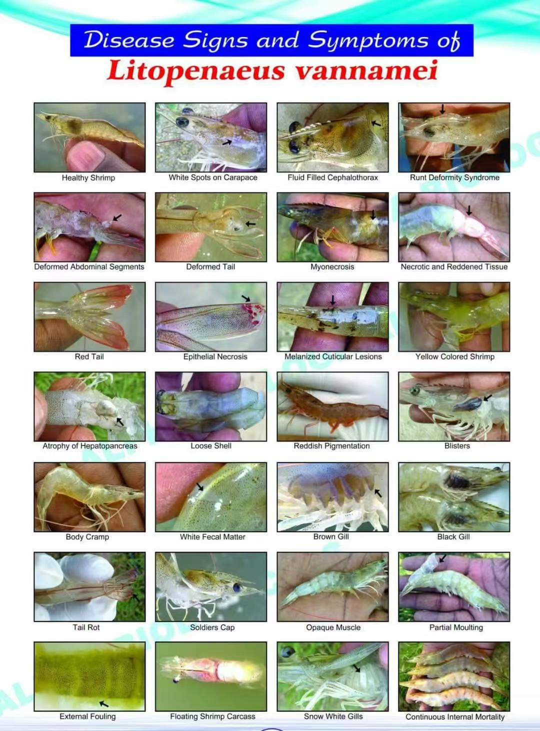
A sign is a reason to test, not a diagnosis
White spots on the carapace are the classic sign of white spot syndrome virus, and also of a bacterial infection and of calcium deposits in hard, high-alkalinity water. A red tail is the textbook sign of Taura syndrome, and also of a shrimp stressed by low oxygen. Mixed infections are common: a pond weakened by EHP is the pond where Vibrio takes hold.
The decision that follows depends on the cause, not the sign. White spot in a grow-out pond means preparing an emergency harvest within days; a bacterial shell infection means fixing the water and the bottom. So the sign chooses the test, and the test chooses the action.
The 28 signs and what to test for
Read across: the sign as it appears on the chart, the causes it is most often associated with, and the PCR kit in this catalogue that confirms or rules out each cause. Where a sign has no single cause, the table says so and points to the core panel.
| Sign on the chart | Most often associated with | PCR to run |
|---|---|---|
| Healthy shrimp | Reference animal: transparent body, firm shell, full gut line | Routine screening: core panel AQ-48 (AHPND, EHP, WSSV, IHHNV) |
| White spots on carapace | White spot syndrome virus (WSSV); also bacterial white spot and mineral deposits in high-alkalinity water | WSSV: AQ-11 · WSSV + DIV1: AQ-22 |
| Fluid filled cephalothorax | No single cause; part of the IHHNV runt deformity syndrome ('bubble head') and of systemic infections | Core panel AQ-48 (includes IHHNV) |
| Runt deformity syndrome | IHHNV, the classic cause of runt deformity syndrome; EHP also stunts growth and widens size variation | IHHNV: AQ-14 · EHP: AQ-01 |
| Deformed abdominal segments | IHHNV (runt deformity syndrome); nutrition and handling damage look similar | IHHNV: AQ-14 |
| Deformed tail | IHHNV: a deformed sixth abdominal segment in runt deformity syndrome, usually with a bent rostrum; also healed injury or bacterial tail necrosis | IHHNV: AQ-14 · Vibrio: AQ-27 |
| Myonecrosis | Infectious myonecrosis virus (IMNV); covert mortality nodavirus (CMNV), Penaeus vannamei nodavirus and microsporidian infection look the same; stress causes a similar necrosis | IMNV: AQ-20 · CMNV + IMNV: AQ-24 |
| Necrotic and reddened tissue | IMNV: white necrosis of the distal abdominal muscle and tail that turns red; CMNV looks similar; Vibrio invades the damaged tissue; acute Taura syndrome virus (TSV) reddens the whole body | CMNV + IMNV: AQ-24 · Vibrio: AQ-27 · TSV: AQ-08 |
| Red tail | TSV in the acute phase, the original 'red tail disease'; also stress | TSV: AQ-08 |
| Epithelial necrosis | Most typical of acute TSV: focal necrosis at the edges of the uropods and pleopods, with a red tail fan; bacterial shell necrosis looks similar | TSV: AQ-08 · Vibrio: AQ-27 |
| Melanized cuticular lesions | TSV in the transition phase; bacterial shell disease | TSV: AQ-08 · Vibrio: AQ-27 |
| Yellow colored shrimp | A yellow head (cephalothorax) can suggest yellow head virus (YHV) on Asian farms; whole-body yellowing is usually non-infectious, from feed pigments or water colour | YHV: AQ-09 |
| Atrophy of hepatopancreas | AHPND (Vibrio carrying the pirAB toxin genes, also called EMS), mostly in the first 35 days after stocking; necrotising hepatopancreatitis (NHP bacterium), chronic, with a soft shell; hepatopancreatic parvovirus, mainly in other penaeid species. EHP slows growth but usually leaves the organ looking normal | AHPND: AQ-10 · NHP: AQ-51 · HPV: AQ-04 · EHP + AHPND: AQ-21 |
| Loose shell | Loose shell syndrome: poor water quality and hepatopancreas disease, including necrotising hepatopancreatitis; no single agent | NHP: AQ-51 · EHP + AHPND: AQ-21 · Vibrio: AQ-27 |
| Reddish pigmentation | Dying WSSV and TSV shrimp turn red; stress does the same | WSSV: AQ-11 · TSV: AQ-08 |
| Blisters | No single cause | Core panel AQ-48 |
| Body cramp | Muscle cramp: temperature shock, handling or mineral imbalance, not a pathogen | No PCR target: check temperature and minerals |
| White fecal matter | White faeces syndrome, associated with EHP and Vibrio; the worm-like bodies in the faeces are sloughed hepatopancreas tissue, not parasites | EHP + V. parahaemolyticus: AQ-30 · EHP: AQ-01 |
| Brown gill | Gill fouling by organic matter and protozoa on a dirty pond bottom | Bottom and water management first; Vibrio AQ-27 if losses follow |
| Black gill | Melanised gill: Fusarium fungi, bacterial infection, or iron and organic deposits | Fusarium: AQ-06 |
| Tail rot | Bacterial erosion, usually Vibrio, after injury | Vibrio: AQ-27 · V. harveyi + V. parahaemolyticus: AQ-28 |
| Soldiers cap | No single cause | Core panel AQ-48 |
| Opaque muscle | IMNV and CMNV; Penaeus vannamei nodavirus and microsporidian infection look the same; also stress and handling | CMNV + IMNV: AQ-24 |
| Partial moulting | Moult problems from mineral imbalance or low alkalinity; infection makes them worse | No single PCR; core panel AQ-48 if losses rise |
| External fouling | Epibionts such as Zoothamnium, Epistylis and filamentous bacteria on a shell that is not moulting | No PCR target: water quality |
| Floating shrimp carcass | Non-specific: any dead shrimp floats once gas forms. Check dissolved oxygen and hydrogen sulphide first, then WSSV, and AHPND within about 35 days of stocking | Core panel AQ-48 (WSSV, AHPND) · then WSSV + DIV1: AQ-22 |
| Snow white gills | No single cause | Core panel AQ-48 |
| Continuous internal mortality | Steady losses on the pond bottom: covert mortality nodavirus (CMNV), chronic IMNV, necrotising hepatopancreatitis, EHP-weakened stock, Vibrio | CMNV + IMNV: AQ-24 · NHP: AQ-51 · EHP + AHPND: AQ-21 |
Which tissue to send for which test
Sample live, moribund animals from the pond edge rather than dead ones from the bottom: the tissue is fresher and the pathogen load is highest. To diagnose a sick pond, 10 to 20 moribund animals are enough. To screen healthy stock or certify post-larvae, test about 150 animals, which detects a 2 percent prevalence with 95 percent confidence, in several small pools rather than one.
Gill and pleopods (swimming legs) for WSSV, IHHNV and TSV; gill, lymphoid organ or haemolymph for YHV. Hepatopancreas for AHPND, necrotising hepatopancreatitis, EHP and hepatopancreatic parvovirus, plus faeces for EHP in broodstock, so the animal stays alive. Tail muscle for IMNV and CMNV. Whole post-larvae, ground together, for a batch test before stocking.
When the signs overlap
Most ponds show more than one sign at once, and the costliest pathogens often travel together. A multiplex kit answers several questions in one tube: the core panel AQ-48 covers AHPND, EHP, WSSV and IHHNV, four of the pathogens farms screen most often; duplex kits pair the causes that share a sign, such as CMNV with IMNV for white muscle, or EHP with Vibrio parahaemolyticus for white faeces.
The detection kits in the aquaculture line are lyophilised, and with the 24-test kits the compact analyser returns the result at the farm about 50 minutes after sampling. The time between seeing a sign and knowing its cause drops from days to an afternoon, which is the window in which the decision still saves the crop.
Written by the Medicare Vet product team from manufacturer data, WOAH guidance and published validation. Not a substitute for veterinary or laboratory advice in your market.
Products discussed
Catalogue items that apply to this note, each with its own page, specifications and quotation form. The full line is on the aquaculture pcr page.
- EMS/EHP-18S/WSSV/IHHNV Nucleic Acid Detection Kit (Real-time PCR, lyophilised)AQ-48 · EMS/EHP-18S/WSSV/IHHNV
- Nucleic Acid Detection Kit for White Spot Syndrome Virus (WSSV) (qPCR Method)AQ-11 · WSSV Nucleic Acid Detection Kit (qPCR Method)
- Duplex Nucleic Acid Detection Kit for White Spot Syndrome Virus (WSSV) and Decapod iridescent virus 1 (DIV1) (qPCR Method)AQ-22 · WSSV & DIV1 Duplex Nucleic Acid Detection Kit (qPCR Method)
- Nucleic Acid Detection Kit for Infectious Hypodermal and Hematopoietic Necrosis Virus (IHHNV)(qPCR Method)AQ-14 · IHHNV Nucleic Acid Detection Kit (qPCR Method)
- Nucleic Acid Detection Kit for Taura Syndrome Virus (TSV) (qRT-PCR Method)AQ-08 · TSV Nucleic Acid Detection Kit (qRT-PCR Method)
- Duplex Nucleic Acid Detection Kit for covert mortality nodavirus (CMNV) & Infectious Myonecrosis Virus (IMNV)(qRT-PCR Method)AQ-24 · CMNV & IMNV Duplex Nucleic Acid Detection Kit (qRT-PCR Method)
- Nucleic Acid Detection Kit for Acute Hepatopancreatic Necrosis Disease (AHPND)(qPCR Method)AQ-10 · AHPND Nucleic Acid Detection Kit (qPCR Method)
- Necrotising Hepatopancreatitis Bacterium (NHPB) Nucleic Acid Detection Kit (Real-time PCR, lyophilised)AQ-51 · NHPB
- Duplex Nucleic Acid Detection Kit for Enterocytozoon hepatopenaei (EHP) and Acute Hepatopancreatic Necrosis Disease (AHPND)(qPCR Method)AQ-21 · EHP & AHPND Duplex Nucleic Acid Detection Kit (qPCR Method)
- Duplex Nucleic Acid Detection Kit for Enterocytozoon hepatopenaei (EHP) and Vibrio parahaemolyticus (Vp) (qPCR Method)AQ-30 · EHP & Vp Duplex Nucleic Acid Detection Kit
- Nucleic Acid Detection Kit for Yellow Head Virus (YHV) (qRT-PCR Method)AQ-09 · YHV Nucleic Acid Detection Kit (qRT-PCR Method)
- Nucleic Acid Detection Kit for Fusarium spp. (Fus) (qPCR Method)AQ-06 · Fus
- Duplex Nucleic Acid Detection Kit for Total Vibrio Count (TVC) and Total Bacterial Count (TBC) (qPCR Method)AQ-27 · TVC & TBC Duplex Nucleic Acid Detection Kit (qPCR Method)
- Compact real-time PCR analyser, 24-wellINS-MG242 · Micgene 242
Questions
Can shrimp disease be diagnosed from a photo?
No. A photo or a chart narrows the list, but several pathogens produce the same sign, mixed infections are common, and stress mimics several of them. The sign tells you which PCR to run; the PCR tells you what to do.
What causes white spots on shrimp?
White spot syndrome virus is the cause to rule out first, because an outbreak can empty a pond within a week. Bacterial white spot and calcium deposits in hard water look similar. A negative WSSV PCR on moribund animals points away from the virus and towards bacterial or mineral causes; if losses continue, resample and test again.
Which single test covers the most signs?
A multiplex panel. AQ-48 tests for AHPND, EHP, WSSV and IHHNV in one tube, four of the pathogens farms screen most often for stunting and mortality in whiteleg shrimp. Add TSV for red tail and IMNV with CMNV for white muscle.
More from the blog
- Isothermal amplification vs lateral-flow vs laboratory PCR: choosing the right test for infectious disease in dogs and cats2026-09-22 · Isothermal amplification
- Quantitative immunofluorescence vs rapid tests for CRP and SAA: why a number changes case management2026-09-22 · Immunofluorescence
- Lyophilised qPCR vs wet-reagent PCR and LAMP for shrimp pathogen screening on the farm2026-09-22 · Aquaculture PCR
- Multiplex real-time PCR panels vs single-target tests: syndromic testing in the veterinary laboratory2026-09-22 · Real-time PCR
- EHP in shrimp: why the hepatopancreas looks normal until it is too late2026-09-21 · Aquaculture PCR
- White spot syndrome virus: three to ten days from first sign to empty pond2026-09-21 · Aquaculture PCR
- Chairside molecular testing: what changes when PCR-grade results arrive in 30 minutes2026-09-21 · Isothermal amplification
- How to read a veterinary rapid test correctly, and when not to trust it2026-09-21 · Rapid test